

Motor disability changes how a person moves through the world, and it quietly changes what happens inside the skeleton too. Picture someone like Daniel, a 52-year-old in Torrance who has used a wheelchair since a spinal cord injury in his twenties. He kept up with therapy, ate well, and felt strong in his arms and shoulders. Then one ordinary transfer from his chair to the couch ended with a broken thigh bone. Nobody had ever checked his bone density.
Stories like this are far more common than most families realize. When muscles can’t pull on bones the way they normally do, the skeleton loses the signals that keep it dense and strong. That loss happens without pain or warning. Over the years, it can turn small bumps, twists, and transfers into serious fractures.
Here’s the hopeful part. Bone loss can be spotted early, long before anything breaks. The tricky part, for anyone living with limited mobility, has always been the testing itself. Getting to an imaging center, sliding onto a narrow table, and holding a fixed position for an X-ray-based scan is a lot to ask.
That’s exactly where a newer, radiation-free approach makes a real difference. This guide walks through why bone health deserves extra attention with a motor disability, what the research actually shows, and how an in-home REMS scan can help people and caregivers stay a step ahead.
Why Motor Disability Puts Bones at Higher Risk
Bone is living tissue. It’s constantly being broken down and rebuilt, and it takes its cues from the forces it feels every day. Walking, standing, climbing stairs, and even the steady pull of working muscles all tell bones to stay strong.
The “Use It or Lose It” Rule of Bone
Scientists have known for more than a century that bone adapts to the load placed on it, a principle often called Wolff’s law. When that load drops, the body starts treating bone as something it doesn’t need as much. Cells that remove bone get busier than the cells that build it, and density slowly slips away.
With a motor disability, the legs and hips often carry far less weight than they used to, or they may never have carried full weight at all. Muscles that are weak, paralyzed, or stiff from spasticity also pull on bone differently. The result is a skeleton that gets thinner and more fragile in the places that matter most for fractures, especially the hip, the knee area, and the lower leg.
Other Factors That Quietly Add Up
Reduced movement is the biggest driver, but it rarely acts alone. Several other things can stack on top of it:
- Certain medications, such as long-term steroids and some anti-seizure drugs, can affect bone turnover or vitamin D levels.
- Less time outdoors can mean lower vitamin D, which the body needs to absorb calcium.
- Swallowing or feeding challenges can make it hard to get enough calcium and protein.
- Hormonal changes, including menopause, speed up bone loss for many women.
- A higher risk of falls during transfers raises the odds that weak bone will actually break.
What the Research Says About Motor Disability and Bone Loss
This isn’t a hunch. Bone loss tied to immobility has been studied for decades, and the findings point in the same direction again and again.
Some of the clearest evidence actually comes from space. Astronauts who spend months in microgravity lose bone in their hips and spine because their legs no longer carry weight. NASA has tracked this closely for years, and it’s one reason astronauts follow strict exercise routines in orbit. The same basic process plays out on Earth when a motor disability takes weight off the legs.
Bed rest studies tell a similar story. Healthy volunteers who stay in bed for several weeks start losing bone at the hip and heel surprisingly fast. Their bodies respond to less load by removing bone that seems unnecessary.
Research in people with a motor disability adds another layer. The National Institutes of Health lists limited activity and long-term use of certain medications among the key risk factors for osteoporosis. Studies in spinal cord injury, cerebral palsy, stroke, and multiple sclerosis all report lower bone density and higher fracture rates compared with the general population.
The takeaway is simple. Less movement usually means weaker bones over time, and the people who face that risk deserve regular, accessible testing.
Common Types of Motor Disability Linked to Bone Loss
A motor disability can come from many different causes, and each one affects bone in its own way. Here are some of the most common.
Spinal Cord Injury
Bone loss after a spinal cord injury can be dramatic, especially in the first year or two. Research consistently shows rapid loss of bone below the level of injury, with the knee region and lower legs hit hardest. Fractures around the knee during routine transfers or minor falls are a well-known problem in this group. For anyone living with this type of motor disability, an early baseline scan is one of the smartest moves they can make.
Cerebral Palsy
Many children and adults with cerebral palsy, particularly those who don’t walk independently, have low bone density. Studies of adults with cerebral palsy have found higher rates of osteoporosis and fractures than in the general population, often at younger ages than doctors would expect.
Stroke
Stroke is one of the leading causes of long-term motor disability in American adults. After a stroke, the weaker side of the body tends to lose bone faster. Combine that with balance problems and a higher fall risk, and it’s easy to see why hip fractures are a concern for stroke survivors.
Parkinson’s Disease and Multiple Sclerosis
Both conditions often lead to a progressive motor disability, and both are linked to lower bone density and more fractures. Reduced activity, medication effects, and frequent falls all play a part.
Muscular Dystrophy and Other Neuromuscular Conditions
Progressive muscle weakness, steroid treatment, and reduced walking can all chip away at bone strength over time. Because this kind of motor disability often begins in childhood, bone checks are worth starting early.
Why Bone Testing Often Gets Skipped
Here’s a frustrating reality. People living with a motor disability are among those who would benefit most from regular bone checks, yet many never get one.
Part of the reason is simple logistics. Getting to an imaging center may mean booking accessible transport, arranging a caregiver, and planning a whole day around a short appointment. Once there, the traditional DXA scan requires lying flat on a narrow table in a fairly specific position. For someone with contractures, scoliosis, spasticity, or pain, that position can be uncomfortable or just not possible.
There’s also an accuracy issue. DXA readings of the spine can look falsely healthy when there’s arthritis, curvature, or calcium deposits in the area, and metal hardware such as rods or hip implants can make some regions impossible to read. For many people with a motor disability, those are exactly the complications they live with.
So the scan gets postponed. Then postponed again. And too often, the first sign of a bone problem is a fracture. That gap is exactly what REMS technology was built to close.
The Real Cost of a Missed Diagnosis
A fracture is painful for anyone. For someone with a motor disability, it can set off a chain reaction that lasts for months.
Healing often takes longer when blood flow and muscle activity are reduced. Casts and braces can cause pressure sores on skin that already has limited feeling. Hospital stays bring infection risks. Time away from therapy can mean losing strength and independence that took years to build. For caregivers, the extra lifting, wound care, and appointments add real physical and emotional strain.
There’s a quieter problem too. Some people with a motor disability have reduced sensation in their legs, so a fracture can go unnoticed for days. Swelling, warmth, or a change in muscle spasms may be the only clue. By the time it’s found, healing is already off to a slow start.
Every one of those outcomes is easier to prevent than to treat. A simple scan that flags weak bone early gives families time to act with exercise, nutrition, safer transfers, and a conversation with their doctor.
What Is REMS Technology?
REMS stands for Radiofrequency Echographic Multi Spectrometry. It’s an ultrasound-based method for measuring bone health at the two sites doctors care about most when it comes to osteoporosis: the lumbar spine and the hip (femoral neck).
Instead of passing X-rays through the body, REMS technology sends harmless ultrasound waves into the bone and analyzes the signals that bounce back. Advanced software compares those signals against reference models of healthy and osteoporotic bone. The scan then produces the familiar numbers doctors already use, including bone mineral density, T-score, and Z-score.
REMS has been cleared by the FDA in the United States and is backed by a growing body of clinical research. A large multicenter study published in Osteoporosis International in 2019 found strong agreement between REMS and DXA when diagnosing osteoporosis at both the spine and the hip. Researchers have also reported that REMS is less affected by some of the artifacts that can throw off DXA readings.
5 Ways REMS Technology Helps People With a Motor Disability
This is where REMS technology really earns its place. For someone with limited mobility, the benefits go well beyond convenience.
1. No Radiation, Ever
REMS technology uses ultrasound, not X-rays. That means zero ionizing radiation for the person being scanned. It’s a meaningful advantage for anyone who may need repeat scans over the years, and it gives families peace of mind.
2. The Scan Comes to You
Superior Bone Scanning brings portable REMS equipment straight to homes and offices across Los Angeles. No accessible van to book. No waiting room. No long day out. For someone living with a motor disability, being scanned in a familiar space, with a caregiver nearby, can turn a stressful appointment into a calm one.
3. Clearer Results When the Spine Is Complicated
Because REMS technology analyzes the full ultrasound signal rather than a single X-ray image, research suggests it’s less likely to be fooled by arthritis, calcifications, or other changes that can inflate DXA spine readings. That matters a lot for people whose bodies don’t fit the textbook picture.
4. A Look at Bone Quality, Not Just Density
Density is only part of the story. REMS technology can also provide a Fragility Score, which gives insight into bone quality and how likely bone is to break, independent of density alone. For someone with a motor disability, where fracture risk is the real concern, that extra layer of information is valuable.
5. Easy Tracking Over Time
Since there’s no radiation and the scan happens at home, it’s much easier to check bone health again after six months or a year. That makes it possible to see whether an exercise or nutrition plan is actually working, rather than guessing.
What to Expect During an In-Home REMS Scan
A lot of people worry the process will be complicated. It isn’t, and every step is designed with motor disability in mind.
A trained technician arrives with portable REMS technology: a compact device and a handheld probe. The person being scanned lies comfortably on their back, on a bed, a couch, or a treatment table. A little warm gel goes on the skin, and the probe is placed gently over the abdomen to scan the lower spine and then over the hip area.
There’s no squeezing, no loud machinery, and no needles. Most people barely feel a thing. The scan is quick, and results are reviewed and explained in plain language, with a clear report you can share with your doctor.
Caregivers are welcome to stay in the room and help with positioning. If someone can’t lie fully flat or has hardware on one side, the technician can talk through options before starting.
Understanding Your REMS Technology Results
A bone density report can look like a page of numbers at first. Here’s what the key ones mean.
T-Score
The T-score compares your bone density with that of a healthy young adult. According to the World Health Organization’s long-standing criteria, a score of -1.0 or higher is considered normal. A score between -1.0 and -2.5 points to low bone mass, often called osteopenia. A score of -2.5 or lower falls in the osteoporosis range.
Z-Score
The Z-score compares your bone density with people of the same age and sex. It’s especially useful for younger adults and for anyone with a motor disability that started early in life, such as cerebral palsy. A low Z-score can signal that something beyond normal aging is affecting bone.
Fragility Score
This is where REMS technology adds something extra. The Fragility Score looks at the quality and structure of bone, not just how dense it is. Two people can have similar density numbers but very different fracture risks. The Fragility Score helps tell them apart.
Your results come with a plain-language explanation, so you’re never left guessing about what the numbers mean for daily life.
Building a Bone Health Plan Around a Motor Disability
A scan is the starting point, not the finish line. Once you know where bone health stands, the next step is doing something about it.
At Superior Bone Scanning, results guide a personalized plan built around exercise and supplements. These are focused, non-drug strategies designed to support long-term bone health, and they work best in coordination with your physician. Follow-up scans with REMS technology then show whether the plan is making a real difference.
Movement That Fits Your Body
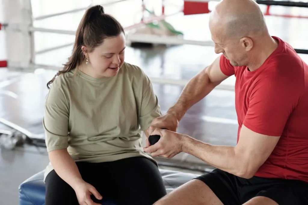
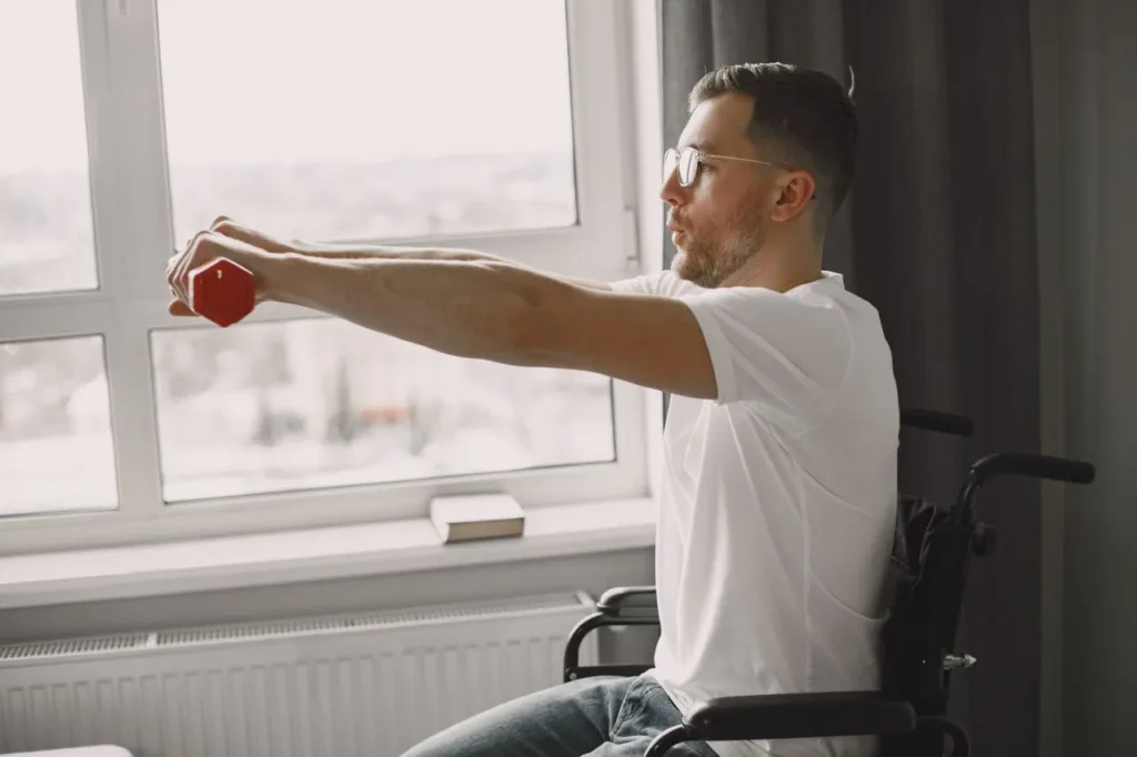
Even with a motor disability, there are often safe ways to load bone. Depending on the person, that might include supported standing in a standing frame, resistance work for the arms and upper body, aquatic therapy, or functional electrical stimulation cycling, which has shown promise in research for people with spinal cord injury. Physical therapists can help tailor what’s realistic and safe.
Smart Nutrition and Supplements
Calcium, vitamin D, protein, and magnesium all play a role in bone health. Many people with a motor disability fall short on vitamin D in particular. Testing helps show whether supplementation makes sense and at what level, rather than taking a one-size-fits-all approach.
Fall and Fracture Prevention
Safer transfer techniques, the right equipment, good lighting, and removing tripping hazards can all lower the chance that weak bone ever gets tested by a fall.
Tips for Caregivers Preparing for a Home Scan
A little planning makes scan day smooth and stress-free. Here are a few things that help:
- Choose a spot where the person can lie on their back comfortably, such as a bed, sofa, or therapy table.
- Wear loose clothing that makes the lower belly and hip easy to reach. No need to remove everything.
- Have a list of current medications and any past fractures handy.
- Let the team know about rods, plates, joint replacements, or a pump ahead of time.
- Plan for any pillows, wedges, or positioning aids the person normally uses.
- Write down questions you’d like to ask after the results come in.
That’s really all it takes. The technician handles the rest and works around the person’s comfort, not the other way around.
REMS Technology for Care Homes, Clinics, and Group Settings
Many people with a motor disability live in assisted living, group homes, or skilled nursing settings. Moving several residents to an outside imaging center is a big lift for staff and a tiring trip for residents.
Superior Bone Scanning can bring REMS technology on site and scan multiple residents in a single visit. Staff stay on their regular schedule, residents stay comfortable, and the facility gets clear bone health data to share with physicians. It’s a practical way to add fracture prevention to a resident care plan without the usual hassle.
Who Should Consider a Bone Scan?
A bone density check is worth discussing with your care team if you or a loved one:
- Has a spinal cord injury, cerebral palsy, multiple sclerosis, Parkinson’s disease, or a history of stroke
- Uses a wheelchair or walks with limited weight bearing
- Has taken steroids or anti-seizure medication for a long time
- Has broken a bone from a minor fall or a routine transfer
- Is a woman going through or past menopause
- Has never had a bone density test at all
If any of these sound familiar, earlier is better. Bone loss is far easier to manage when it’s caught before a fracture, and REMS technology makes that first test simpler than ever.
Conclusion
Living with a motor disability already means managing plenty. Bone health shouldn’t be one more thing left to chance, especially when silent bone loss can lead to painful, life-changing fractures.
The research shows that reduced movement puts bones at risk. The encouraging news is that early detection is now easier, safer, and more comfortable than ever. REMS technology offers accurate, radiation-free testing that comes straight to your door, followed by a personalized exercise and supplement plan designed around your body and your goals.
Whether you’re living with a motor disability yourself or caring for someone who is, the best time to check bone health is before a fracture forces the question.
Ready to take the first step? Book your in-home REMS bone scan with Superior Bone Scanning and get clear answers about bone health from the comfort of home.
Frequently Asked Questions
Does a motor disability always lead to bone loss?
Not always, but the risk is clearly higher. Less weight-bearing and reduced muscle pull mean bones get fewer signals to stay strong.
Is REMS technology safe for people with limited mobility?
Yes. It uses ultrasound instead of radiation, and the scan is done lying comfortably at home.
How accurate is a REMS scan compared with DXA?
Clinical studies show strong agreement between REMS and DXA for diagnosing osteoporosis at the spine and hip. REMS may also be less affected by arthritis and calcifications.
Can someone with a motor disability be scanned at home?
Absolutely. Superior Bone Scanning brings portable equipment to homes, care facilities, and offices across Los Angeles.
How often should bone density be checked with a motor disability?
That depends on your health history and your doctor’s advice. Because REMS is radiation-free, repeat scans every 6 to 12 months are easy to plan if needed.
Does REMS technology work if I have metal implants?
Often, yes. The technician can review any hardware beforehand and focus on the sites that give the clearest results.