

REMS imaging is changing what a bone density scan actually feels like to get. No referral chain to untangle, no radiology department across town, no lead apron, no machine that hasn’t changed much in thirty years. It reads bone density using ultrasound instead of X-rays, covers both scan sites in well under ten minutes, and hands back a result the same day, often within minutes of the appointment ending. This piece walks through what’s actually happening inside the machine, what the research says about how well it performs, and why a technology that barely existed in U.S. clinics a few years ago now has its own federal billing code.
What Is REMS Imaging, Exactly?
REMS stands for Radiofrequency Echographic Multi Spectrometry, which is a mouthful even for the people who use it every day, so it usually just gets called REMS imaging. It’s a bone density scan built around ultrasound rather than radiation, and it reads the two sites that matter most for fracture risk: the lumbar spine and the femoral neck, the top of the thighbone where it meets the hip. Those are the same two sites a DEXA scan has always targeted, which is part of why the two technologies compare so directly.
What makes this different from a routine ultrasound is what the machine does with the signal after it bounces back off bone. A standard ultrasound compresses raw sound data down into a picture and discards most of the underlying signal in the process, since a picture is all it needs to produce. This approach keeps that raw radiofrequency signal completely intact and studies its full spectrum instead, which is where the “multispectrum” part of the name comes from. That untouched signal carries a level of density and microstructure detail a picture was never designed to hold onto, and it’s the reason the resulting readout can pick up nuances a conventional scan simply misses.
The Research Behind How REMS Imaging Reads Bone
Here’s the part that actually explains why this works. Every bone reflects an ultrasound signal in a slightly different way depending on its density, its internal architecture, and its composition. Researchers built a large reference library from thousands of earlier scans, some from people with strong, healthy bone and some from people with confirmed osteoporosis, and mapped the spectral pattern typical of each group.
During the appointment itself, the device compares a patient’s live signal against that reference library, adjusting for age, sex, BMI, and which skeletal site is being scanned. A closer match to a healthy pattern produces a higher density score. The system also automatically screens out unreliable readings, things like calcifications, arthritic changes, or soft tissue interference, so a single stray data point doesn’t throw off the whole result. That built-in filtering is a big reason the same patient tends to get consistent readings across visits, regardless of which technician runs the scan, and it’s part of what makes the technology dependable enough for repeat monitoring over time rather than a one-off check.
A lumbar spine pass takes roughly 80 seconds, and a femoral neck pass around 40 seconds, so a full REMS imaging bone density scan covering both sites usually wraps up in well under ten minutes once someone is positioned and settled. There’s no separate machine to move to for the second site. One handheld probe covers both.
Walking Through an Appointment
Part of why this bone density scan has caught on so quickly is how uneventful the actual appointment is. No fasting beforehand, no contrast dye, no gown, nothing to remove except jewelry sitting directly over the scan area. A technician applies a small amount of gel to the lower back and hip, moves a handheld probe slowly across each site, and the device quietly gathers thousands of individual signal readings per location while that’s happening.
Because the whole process runs on sound waves instead of X-rays, there’s no radiation dose to log, no lead-lined room required, and nothing keeping a provider from setting up the equipment somewhere other than a hospital basement. That single fact is what makes a mobile bone density scan possible at all. A DEXA machine is large, fixed equipment permanently installed inside a shielded room. REMS equipment is compact enough to travel to a home, an office, or a community wellness event, which is exactly how Superior Bone Scanning delivers this kind of appointment across Los Angeles instead of asking people to come find one.
Results are typically ready the same day, and often within minutes of the scan finishing, since there’s no film to develop and no separate radiology read sitting between the appointment and the report.
What the Report Actually Tells You
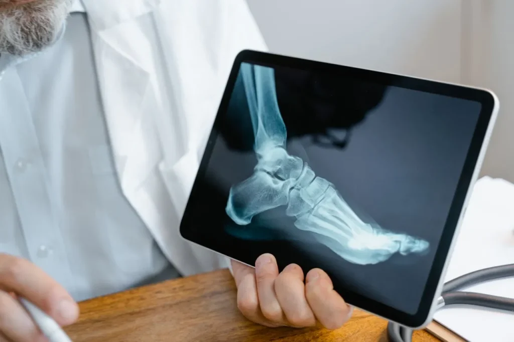
The output will look familiar to anyone who has had a bone density scan before. A T-score compares bone density to that of a healthy young adult, and a Z-score compares it to people of the same age and sex. Those are the same two figures a physician already uses to sort results into normal, osteopenic, or osteoporotic, and they sit on the exact same scale DEXA uses, so nothing new has to be translated or relearned.
There’s also a number DEXA doesn’t produce: the Fragility Score. Instead of measuring density on its own, it evaluates bone microstructure and estimates fracture likelihood over roughly the next five years, independent of the density figure by itself. Two people can post nearly identical T-scores and still carry meaningfully different real-world fracture risk, and the Fragility Score exists specifically to catch that gap.
REMS Imaging Now Has Its Own U.S. Billing Code
For years, one of the biggest practical hurdles for this technology in the United States wasn’t accuracy. It was billing. As of January 2024, REMS bone assessment carries its own Category III CPT code, 0815T, officially described as ultrasound-based radiofrequency ultrasound multispectrometry for bone density and fracture-risk assessment at the hip, pelvis, or spine. That single change matters more than it sounds like it should. A recognized billing code is what lets providers submit claims consistently and gives insurers a defined procedure to evaluate, rather than treating every scan like this as an unclassified service.
It doesn’t guarantee coverage on every plan, and it’s worth checking directly with an insurer or with Superior Bone Scanning about current options before assuming either way. But it’s a meaningful signal that this bone density scan is moving from a niche offering toward standard practice in U.S. clinical settings, and it’s part of why more providers are adding it each year.
REMS Imaging vs. DEXA: What the Evidence Shows
DEXA remains the long-standing standard for a bone density scan, and nothing here argues it should be replaced. But REMS imaging has built a genuine research base since receiving regulatory clearance. A review published in Aging Clinical and Experimental Research, produced by an expert consensus group convened by the European Society for Clinical and Economic Aspects of Osteoporosis, found sensitivity and specificity both above 90% when measured against DEXA as the reference standard. That’s a strong showing for a method that involves no radiation at all.
Precision is where the newer technology has a real edge. Repeatability studies have measured a precision error around 0.38% at the lumbar spine, a figure that compares favorably to typical DEXA benchmarks and points to less variation between repeat scans on the same person, whether the same technician runs both or two different ones do. For anyone tracking bone density over months or years of treatment, that consistency is what turns a follow-up scan into something genuinely useful instead of statistical noise.
A 2025 case series in BMC Medical Imaging also looked at how REMS performs in more complicated situations, including patients being monitored for rare bone disorders, and found it held up as a workable option where traditional imaging can be harder to interpret. Separate 2025 research has specifically examined bone density readings in patients before and after total hip replacement, an area where DEXA has always struggled because metal hardware distorts an X-ray-based reading.
A separate real-world validation study out of Australia has also looked at how the lumbar spine and hip readings hold up outside a controlled research setting, the kind of everyday clinical use that matters more to patients than a lab environment ever could. It’s worth being upfront that this remains an active area of study, and not every question about how demographic inputs shape the final reading has been fully settled. For most people simply asking whether the result can be trusted, the honest answer is that REMS imaging performs well against DEXA on classification accuracy while offering a very different, and for many people far more accessible, path to get there.
Who Tends to Benefit Most
Skipping radiation opens the door for people DEXA has always been complicated for. Pregnant women generally can’t get a DEXA scan at all because of the radiation exposure, but this kind of bone density scan carries no such restriction, which makes it a practical option for anyone who needs bone health monitored during pregnancy or shortly after.
People with metal implants, spinal hardware, or joint replacements also tend to get cleaner readings, since the technology isn’t interpreting density through X-ray attenuation the way DEXA is, and metal doesn’t distort an ultrasound signal the way it distorts an X-ray image. Athletes and highly active adults who put unusual, repetitive stress on their joints are another group worth mentioning, since a baseline reading early on gives a physician something concrete to compare against if a stress fracture or unexplained pain ever shows up later.
Younger adults who want a baseline before anything changes, people managing thyroid conditions that affect bone turnover, and anyone already on osteoporosis medication who wants proof it’s actually working are strong candidates too. Because the scan travels to the patient rather than the reverse, it also removes one of the biggest everyday barriers to getting screened in the first place: finding the time to get to an imaging center, sit in a waiting room, and get back to the rest of the day.
Even NASA Is Paying Attention
One of the more unexpected signals of confidence in this technology came from outside the clinic entirely. Echolight, the manufacturer behind the equipment, has fitted NASA-affiliated spaceflight simulation research with its devices to monitor bone health, since astronauts lose bone density rapidly in microgravity and need frequent, portable, radiation-free monitoring that doesn’t add to their overall radiation exposure. It’s a niche application, but it says something concrete about how much confidence researchers place in the accuracy of a scan like this when the margin for error actually matters.
REMS Imaging Near You: What Mobile Actually Means
The near-me search that usually leads someone to this technology tends to start from a specific moment: a doctor mentioned bone density in passing, or a friend was just diagnosed with osteoporosis, and getting checked suddenly feels urgent. Superior Bone Scanning built its Los Angeles service around exactly that moment. Rather than a single fixed office, the portable equipment travels to homes, workplaces, senior communities, and scheduled group events across the city, so the closest available appointment is usually the one that comes to a kitchen table rather than the other way around.
That mobility isn’t layered on top of the technology. It’s a direct result of how the scan works. Because there’s no radiation, no shielded room requirement, and no bulky fixed machine, a technician can set up a full appointment almost anywhere with a flat surface and a few quiet minutes. For older adults with mobility limitations, busy parents who can’t carve out half a day, and anyone who has simply never gotten around to booking the traditional version, that difference is often what turns “I should probably get that checked” into an appointment actually on the calendar.
What Happens After Your Results Come Back
A scan only matters if it leads somewhere, and that’s usually where people have the most questions. If the T-score and Z-score land in a normal range, that’s a genuinely reassuring result and a solid baseline to compare against later. If the numbers fall into osteopenic or osteoporotic territory, the next step isn’t automatically a prescription. Superior Bone Scanning builds a plan around targeted exercise and supplementation first, aimed at supporting bone density naturally, and treats pharmaceutical treatment as one option among several rather than the default starting point.
That distinction matters because a lot of people who get an unexpected result are simply looking for direction, not a diagnosis to sit with alone. Knowing which exercises actually load bone usefully, which nutrients matter most for the specific site that came back low, and how often to repeat the scan to track change turns a single number into an actual plan. Because the same portable equipment can return for a follow-up appointment, tracking progress over six or twelve months doesn’t mean starting the whole process over.
For people already on osteoporosis medication, that follow-up piece carries extra weight. Bone density can take months to respond to treatment, and without a repeat scan there’s no real way to know whether the current plan is working or whether it’s time to talk to a physician about adjusting it. Because REMS imaging reads with high repeatability between visits, a change in T-score between two appointments is far more likely to reflect a real shift in bone density than measurement noise, which is exactly what makes scheduling that follow-up worthwhile.
There’s also a practical, everyday benefit that tends to get overlooked. Because the appointment itself is short and comes to wherever the patient already is, follow-up scans actually happen on schedule instead of quietly slipping for months at a time. A lot of monitoring plans fail not because the science is wrong but because getting back to an imaging center never quite makes it to the top of the to-do list. Removing that friction is arguably as important to a good outcome as the accuracy of the reading itself.
REMS Imaging Isn’t a Heel Ultrasound
One point worth clearing up directly: this is not the same thing as the quick heel ultrasound sometimes offered at pharmacies or health fairs. A heel ultrasound only estimates density at the heel bone, a site that isn’t especially predictive of hip or spine fracture risk, and it’s generally treated as a rough screening tool rather than a diagnostic one.
REMS imaging reads the lumbar spine and femoral neck directly, the two sites clinicians actually rely on for an osteoporosis diagnosis, and its results land on the same T-score and Z-score scale used with DEXA. If a heel ultrasound at a pharmacy ever flagged a concern, that’s a reasonable prompt to book a more complete bone density scan rather than a diagnosis on its own.
Is REMS Imaging Right for You?
Most adults over 50, anyone with a family history of osteoporosis, postmenopausal women, people on long-term steroid medication, and anyone who has already broken a bone from a minor fall are the groups most commonly advised to get a bone density scan in the first place. None of those risk factors change based on which technology does the reading. What changes is how easy the process is to actually complete, and that’s the piece this technology solves. For someone who has been putting off a scan because of the hassle, the radiation exposure, or simply not having a nearby imaging center, this is very often the version that finally gets booked.
Why Superior Bone Scanning Uses REMS Imaging
Superior Bone Scanning built its entire model around this technology because it matches how people actually want to be cared for. A bone density scan shouldn’t require a half-day off work or a referral chain that takes weeks to untangle. Superior Bone Scanning brings REMS imaging directly to homes, offices, and community events across Los Angeles, and pairs every scan with a personalized plan built around exercise and targeted supplementation rather than defaulting straight to pharmaceutical treatment. For anyone looking for the earliest possible warning sign of bone loss, or who simply wants a starting point they can act on right away, that combination of accessible testing and a genuine follow-up plan is the whole point.
Summary
REMS imaging exists because checking your bone health shouldn’t require radiation, a hospital visit, or weeks of waiting on a referral. It reads the same lumbar spine and femoral neck sites doctors have always relied on; it’s backed by a growing body of published research, and it now carries its own U.S. billing code, a real signal of how far the technology has come. If it’s been years since your last bone density scan, or you’ve never had one, Superior Bone Scanning can bring a REMS imaging appointment to your home or office anywhere in Los Angeles. Book a scan today and get a clear, radiation-free answer about where your bones actually stand.
Frequently Asked Questions
Is REMS imaging as accurate as a DEXA bone density scan?
Research shows REMS imaging classifies osteoporosis with sensitivity and specificity above 90% compared to DEXA, and it holds up well as a reliable bone density scan option.
Does REMS imaging involve any radiation?
No. REMS imaging relies entirely on ultrasound sound waves, so there’s no radiation dose, no lead apron, and no exposure limit to track between scans.
How long does a REMS imaging appointment take?
A full REMS imaging bone density scan covering both the lumbar spine and femoral neck usually finishes in well under ten minutes.
Is there a billing code for REMS imaging in the U.S.?
Yes. REMS imaging received its own CPT code, 0815T, in January 2024, covering bone density and fracture-risk assessment at the hip, pelvis, or spine.
Can I get a REMS imaging scan without a doctor’s referral?
Not with Superior Bone Scanning. You can book a REMS bone density scan directly, and the results can always be shared with your physician afterward.
Is REMS imaging safe during pregnancy?
Yes. Since there’s no radiation involved, a REMS bone density scan is considered safe during pregnancy, unlike a standard DEXA scan.
What does the Fragility Score from a REMS imaging scan measure?
It estimates fracture risk based on bone microstructure, giving a fuller picture than a density number alone can offer.
How soon are REMS imaging results available?
Most people receive their bone density scan results the same day, often within minutes of the scan being completed.