

Reduce fracture risk before a single bad step on a wet sidewalk turns into a hospital stay, and you’re already ahead of where most people start. Bone loss rarely announces itself. It doesn’t ache the way a pulled muscle does, and it doesn’t show up in the mirror the way weight change might. For years, sometimes decades, a skeleton can be quietly losing density while everything on the surface looks completely normal.
Then one ordinary moment, a missed stair, a stumble off a curb, a hard sneeze, changes everything. According to the Bone Health and Osteoporosis Foundation, roughly one in two women and up to one in four men over age 50 will break a bone because of low bone density. Those aren’t small odds, and they’re exactly why learning how to reduce fracture risk has moved from a niche medical topic to something every adult over 50, and plenty of people younger, should understand.
This article walks through what actually drives fracture risk, what the research says about catching it early, and how a new generation of radiation-free scanning technology is changing what “getting checked” even looks like. Whether the goal is protecting personal bone strength, helping an aging parent figure out how to reduce fracture risk, or simply making sense of a confusing lab report, the objective stays the same: know where things stand before biology makes the decision instead.
What “Fracture Risk” Actually Means
Fracture risk is the likelihood that a bone will break under stress that a healthy skeleton would shrug off without issue. It’s not just about whether someone has osteoporosis on paper. Two people can carry the exact same T-score and walk away with very different real-world odds of a break, because fracture risk also depends on bone quality, muscle strength, balance, medication history, and even how someone’s home is laid out. That’s part of why doctors increasingly talk about fracture risk as its own measurable thing, separate from a bone density number alone, and why learning to reduce fracture risk takes more than a single test result.
The most widely used clinical tool for estimating this is FRAX, the Fracture Risk Assessment Tool developed by the World Health Organization, which combines bone density with age, prior fractures, family history, and lifestyle factors to estimate a person’s 10-year probability of a major fracture. It’s a useful starting point, but it was built for population-level screening, not for catching the finer details of bone quality in any one individual. Newer scanning technologies are beginning to build similar predictive scoring directly into the scan itself, which is a meaningful shift in how people reduce fracture risk long before a break ever happens.
It also helps to separate two ideas that get lumped together: fracture risk and osteoporosis. Osteoporosis is a diagnosis, a specific T-score threshold. Fracture risk is a spectrum, and someone can sit in the osteopenia range and still carry meaningfully elevated fracture risk if their bone quality is poor or their balance is unsteady.
Why Bone Strength Matters More Than People Realize
Bone strength isn’t just density. It’s a combination of how much mineral is packed into the bone and how well that bone is structurally organized on the inside. Picture two pieces of wood with the same weight: one solid, one riddled with tiny internal cracks. They’d feel similar in your hand, but one snaps under pressure that the other could easily handle. That’s essentially what happens inside a bone as osteoporosis progresses. Bone strength quietly erodes from the inside while the outer shape looks unchanged, which is exactly why so many fractures come as a total surprise.
Understanding bone strength, not just a single density number, is central to any real plan to reduce fracture risk. A scan that only measures mineral density can miss people whose bone strength has been compromised structurally even though their overall density still looks borderline acceptable. That gap is one of the biggest reasons researchers have pushed for tools that assess bone quality alongside density, not instead of it.
There’s a second layer worth understanding: bone turnover, the ongoing cycle of old bone breaking down and new bone rebuilding. In a healthy adult, this stays roughly balanced. As estrogen and testosterone decline with age, the balance tips toward breakdown, and bone strength erodes even in people whose density score hasn’t dropped into an alarming range yet.
The Numbers Behind Fracture Risk
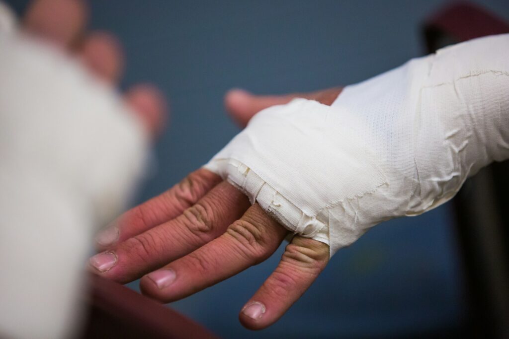
The statistics here are sobering enough to be worth sitting with for a moment. The Bone Health and Osteoporosis Foundation reports that about 10 million Americans currently have osteoporosis, with another 44 million living with low bone density that puts them at meaningfully elevated fracture risk. A hip fracture, the most serious of the common breaks, changes the trajectory of a person’s life more often than most people expect: research summarized by the National Institutes of Health has found that roughly 20 to 24 percent of hip fracture patients over 50 die within a year of the injury, and many who survive never fully regain their previous mobility.
The financial side rarely gets talked about, but it belongs in this conversation too. A single hip fracture often means a hospital stay, surgery, and weeks of rehabilitation, costs that dwarf what a screening and prevention plan would have cost years earlier.
None of this is meant to scare anyone into panic. It’s meant to explain why “wait and see” is rarely the right approach once someone is past 50, has a family history of fractures, or has any of the risk factors below. The first step to reduce fracture risk is knowing where things actually stand, not guessing, and the earlier that starting point is established, the more options stay on the table.
Who’s Most at Risk: A Closer Look
Fracture risk doesn’t affect every group the same way, and painting everyone with the same brush misses some important nuance.
Postmenopausal women carry the highest documented fracture risk, largely because estrogen decline accelerates bone turnover, sometimes causing a woman to lose up to 20 percent of her bone density in the five to seven years following menopause. This is why so much of the existing screening guidance centers on women 65 and older, though anyone with early or surgically induced menopause should be talking about how to reduce fracture risk years sooner.
Men are frequently overlooked here, which is a real problem: roughly one in four men over 50 will experience an osteoporosis-related fracture, and men who fracture a hip actually face a higher one-year mortality rate than women who do. Bone density testing has historically been marketed toward women, leaving a lot of men unaware that fracture risk applies to them too.
People managing chronic conditions like rheumatoid arthritis, celiac disease, or chronic kidney disease also carry disproportionately high fracture risk, both from the disease process itself and from medications like long-term corticosteroids. Cancer survivors on aromatase inhibitors for breast cancer fall into this same higher-risk category and are frequently never told to get a baseline scan.
There’s also a quieter group: people in their 30s and 40s who assume fracture risk is a problem for “later.” Peak bone mass is typically reached by the late 20s, so a baseline scan earlier in adulthood gives someone real information to compare against down the road.
What Determines Your Fracture Risk
A handful of factors show up again and again in the research on who breaks a bone and who doesn’t.
Age and hormonal changes. Bone remodeling naturally tips out of balance with age, and menopause accelerates that shift sharply because estrogen plays a protective role in maintaining bone strength. This is a big part of why postmenopausal women make up such a large share of fracture risk cases, though the underlying hormonal shift affects men too, just more gradually.
Family history. A parent’s hip fracture is one of the strongest predictors of someone’s own risk, independent of their current bone density score. Genetics influence peak bone mass, bone shape, and how quickly bone strength declines with age, which is why family history carries so much weight in a fracture risk assessment.
Medications and medical conditions. Long-term steroid use, some acid reflux medications, certain cancer treatments, thyroid disorders, and chronic kidney disease can all quietly chip away at bone strength as a side effect rather than a primary symptom. Anticonvulsants and certain diabetes medications have also been linked to increased fracture risk in more recent research, which is worth raising with a prescribing physician if it applies.
Lifestyle factors. Smoking, heavy alcohol use, a sedentary routine, and a diet consistently low in calcium, vitamin D, or protein all contribute to weaker bones over time, and changing any one of them is a real way to reduce fracture risk. Even factors like excessive caffeine intake or chronic low body weight have been associated with lower bone density in multiple studies.
Prior fractures. A previous fragility fracture, even a small one in the wrist, roughly doubles the likelihood of a future fracture, which makes early screening after any unexplained break especially important. This is sometimes called the fracture cascade, and it’s one of the clearest signals in all of fracture risk research.
Body frame and mobility. Smaller-framed individuals generally have less bone mass to begin with, and reduced muscle strength or poor balance raises the odds that a stumble turns into a fall, and that a fall turns into a break.
Traditional Bone Scans vs. a Radiation-Free Alternative
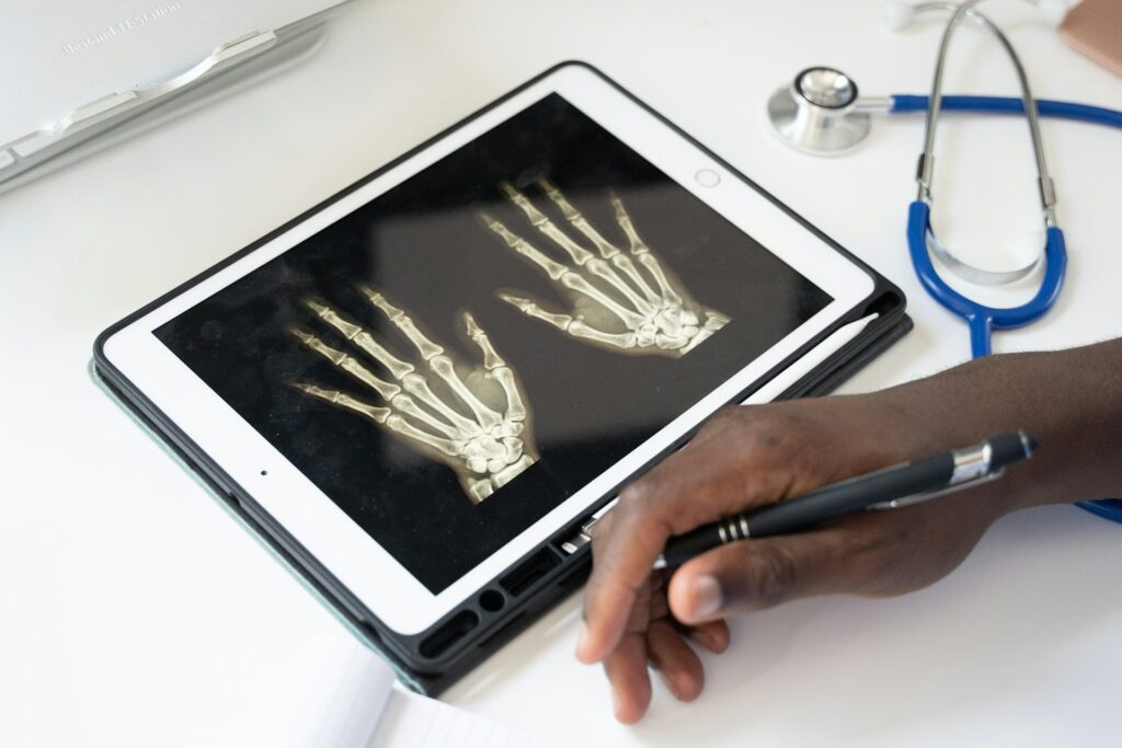
For decades, DEXA scanning has been the standard way to measure bone density, and it still has a legitimate place in bone health care. But it uses ionizing radiation, requires a trip to an imaging center, and can produce misleading readings in people with spinal arthritis, vascular calcification, or metal implants and spinal hardware, all of which can artificially inflate the density number and mask real risk. For pregnant women, and for anyone who wants to reduce fracture risk while keeping radiation exposure low over a lifetime of repeat scans, those limitations matter.
REMS, short for Radiofrequency Echographic Multi Spectrometry, takes a different approach. Instead of X-rays, it uses ultrasound-based radiofrequency signals to evaluate both bone density and bone quality, without exposing the patient to any radiation at all. A multicenter study published in Aging Clinical and Experimental Research found REMS achieved sensitivity and specificity both above 91 percent compared directly against DEXA, with strong statistical agreement between the two methods. That level of agreement matters, because it means switching to a radiation-free option doesn’t mean sacrificing accuracy to gain convenience.
There’s also a practical dimension worth naming. DEXA requires specialized fixed equipment, which is part of why appointments often mean driving across town and sitting in a waiting room. REMS technology is portable by design, which is what makes an in-home or in-office scan possible in the first place, removing a real barrier for older adults, caregivers, and anyone who’s simply put off a scan because it felt like a hassle.
How REMS Technology Helps You Reduce Fracture Risk
What sets REMS apart isn’t just the absence of radiation; it’s what the scan can actually predict. A five-year follow-up study published in the journal Bone found that the REMS-based Fragility Score was a significant independent predictor of incident fragility fractures, meaning the scan doesn’t just describe current bone density; it helps forecast future risk. A separate observational study on the lumbar spine Fragility Score reached similar conclusions, reinforcing that this kind of scoring adds real predictive value beyond a T-score alone.
That distinction matters enormously for anyone trying to reduce fracture risk proactively rather than reactively. A scan that only reports “normal” or “low” density leaves people guessing about what comes next. A scan that also produces a fragility-based risk score gives both the patient and their provider something concrete to act on, whether that means starting a targeted exercise plan, adjusting supplementation, or scheduling closer monitoring.
A REMS scan itself takes only a few minutes. There’s no gown, no changing room, and no lying still under a humming machine arm. A portable ultrasound-style probe passes gently over the hip and lower spine, and the results, including bone density, T-score, and fragility scoring, are typically reviewed the same day in plain language rather than handed over as a printout and a shrug. Patients often say the most valuable part isn’t the technology itself; it’s finally having a real conversation about what their numbers mean and how to reduce fracture risk from there.
Everyday Ways to Reduce Fracture Risk and Build Bone Strength
Genetics and age set the starting line, but daily habits still move the needle more than most people expect, and this is where fracture risk becomes something people can actively influence rather than just monitor.
Weight-bearing exercise. Brisk walking, stair climbing, dancing, and resistance training all send a mechanical signal to bone tissue that says, essentially, keep building. This is one of the single most effective ways to reduce fracture risk at any age, and research shows even adults in their 70s and 80s see measurable gains in bone strength after starting resistance training.
Balance and stability training. Yoga, tai chi, and simple single-leg standing drills won’t add bone density on their own, but they dramatically cut the odds of the fall that turns fragile bone into a fracture in the first place. Fall prevention is, in a very real sense, fracture prevention, and it’s frequently the most underrated piece of any plan to reduce fracture risk.
Calcium and vitamin D. Calcium-rich foods like yogurt, sardines with the bones in, and leafy greens supply the raw mineral bone needs, while vitamin D is what allows the body to actually absorb that calcium. The two work as a pair, and skimping on either one undercuts bone strength over time.
Protein intake. Bone is roughly half protein by volume, and adults who consistently fall short on protein tend to see faster bone loss, particularly after 50.
Cutting back on smoking and heavy drinking. Both habits interfere with the body’s natural bone-building process and are two of the more reversible contributors to declining bone strength.
Home safety adjustments. Securing loose rugs, adding grab bars in bathrooms, and improving hallway lighting won’t change a bone density score, but they remove the everyday hazards most falls actually come from. The Centers for Disease Control and Prevention notes that most falls among older adults happen at home, making this one of the most overlooked pieces of the fracture risk picture.
Regular vision and medication reviews. Poor eyesight and certain medications, particularly sedatives, can affect balance and reaction time. An annual review of both with a physician is a simple, often-skipped way to reduce fracture risk.
When Should You Get Screened?
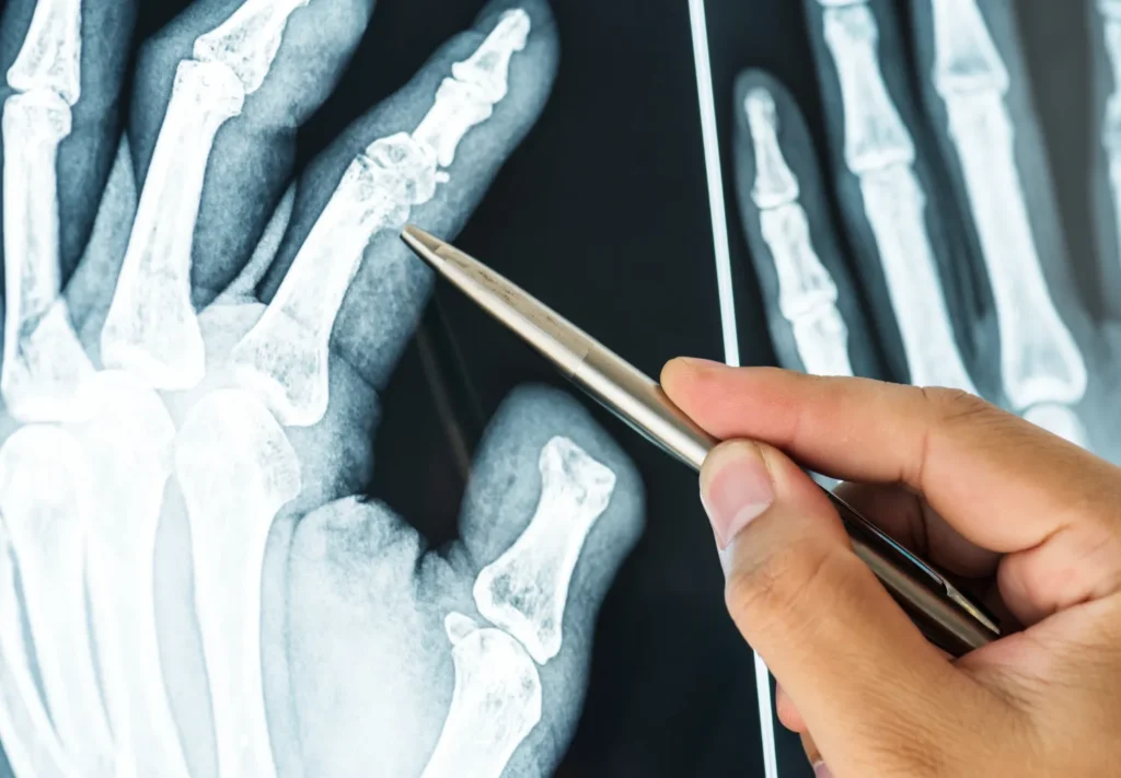
Most guidelines recommend women begin bone density screening at 65, and earlier with risk factors like early menopause, a family history of fractures, or long-term steroid use. Men are generally advised to start around 70, or sooner if risk factors are present. Anyone searching for a bone density scan near me after a first fragility fracture, a worrying family history, or a new diagnosis like rheumatoid arthritis or celiac disease shouldn’t wait for a routine checkup to bring it up. The sooner you get screened, the sooner you can reduce fracture risk with a clear plan.
This is also where accessibility matters more than people expect. A scan that requires a special trip across town, a full afternoon off work, and a cold imaging suite is a scan a lot of busy adults and caregivers keep postponing. As Los Angeles’s most trusted REMS bone scanning provider, Superior Bone Scanning brings the entire process directly to homes and offices throughout the city, whether that’s a condo in Santa Monica, an office in Pasadena, or a family home in the South Bay, which removes one of the biggest reasons people put off finding out where they actually stand.
For caregivers specifically, this convenience solves a real logistical problem. Coordinating a parent’s transportation to a medical imaging center, working around their schedule, and managing the whole outing can turn a fifteen-minute scan into a half-day event. A mobile REMS scan compresses all of that down to a single visit at home, which tends to be the difference between a screening that actually happens and one that keeps getting postponed indefinitely.
What to Expect During and After Your Scan
For anyone who’s never had a bone density scan before, or who’s only had the traditional hospital version, it helps to know exactly what the process looks like. A technician arrives at the scheduled location with a portable, ultrasound-style device; no radiation shielding is required, and no special prep beforehand is needed beyond avoiding calcium supplements for 24 hours if requested. The scan itself involves lying comfortably while the probe passes over the hip and lumbar spine, a process that typically takes just a few minutes per site.
Once the scan is complete, results are generated almost immediately, including a T-score, a comparison to age-matched peers, and, where applicable, a fragility-based fracture risk score. Rather than mailing a report or scheduling a separate follow-up appointment weeks later, results are typically walked through in person, in plain language, right after the scan. That means questions get answered on the spot: what the numbers mean, whether they represent normal bone density, osteopenia, or osteoporosis, and what next steps, if any, make sense.
For many patients, that conversation is the real turning point. A number on a page is easy to file away and forget. A clear explanation of what it means for real-world fracture risk is much harder to ignore, and it’s often the push people need to finally reduce fracture risk.
Summary
The decision to reduce fracture risk isn’t a someday project. It’s a decision that gets easier, cheaper, and far more effective the earlier it’s made. The research is consistent on this point: bone loss is largely silent until a fracture forces the conversation, but a scan that measures both density and bone strength can put real numbers behind a risk that used to be invisible.
Superior Bone Scanning uses advanced REMS technology to bring radiation-free, mobile bone density testing straight to homes and offices across Los Angeles, paired with tailored exercise and supplement guidance built around actual results rather than a generic pamphlet. If protecting bone strength and working to reduce fracture risk has been on the back burner, this is the moment to move it to the front.
Frequently Asked Questions
What does it actually mean to reduce fracture risk?
It means taking steps, through screening, lifestyle changes, and sometimes treatment, to lower the odds that a bone will break under everyday stress. It starts with knowing your current bone density and fragility score, not guessing, and then building a plan around what those numbers actually show.
Can you reduce fracture risk without medication?
Often, yes, especially in the earlier stages of bone loss. Weight-bearing exercise, adequate calcium and vitamin D, balance training, and quitting smoking can meaningfully improve bone strength for many people before medication is ever necessary, though that decision should always involve a physician.
What’s the difference between bone density and bone strength?
Bone density measures how much mineral is packed into bone tissue. Bone strength also accounts for the internal structure and quality of that bone, which is why two people with similar density scores can have different real-world fracture risk.
Is a REMS scan as accurate as a DEXA scan for assessing fracture risk?
Published research shows REMS correlates strongly with DEXA, with sensitivity and specificity both above 90 percent in multiple studies, while also adding fragility-based fracture risk scoring that DEXA doesn’t provide.
How often should someone get rescanned to track fracture risk?
Most providers recommend rescanning every one to two years, though the right interval depends on age, initial results, and whether someone is actively working to reduce fracture risk through treatment or lifestyle changes.
Does a REMS scan hurt or involve radiation?
No. A REMS scan uses ultrasound-based radiofrequency signals instead of X-rays, so there’s no radiation exposure at all, and the scan itself is painless and takes just a few minutes.
Who should prioritize getting screened for fracture risk?
Women 65 and older, men 70 and older, anyone with a prior fragility fracture, a family history of hip fractures, long-term steroid use, or a condition like rheumatoid arthritis or celiac disease should all prioritize screening sooner rather than later.
Does Superior Bone Scanning offer mobile bone density testing in Los Angeles?
Yes. Superior Bone Scanning brings radiation-free REMS testing directly to homes and offices throughout Los Angeles, making it easier for busy adults, seniors, and caregivers to find out where they stand without adding another appointment to an already full schedule.